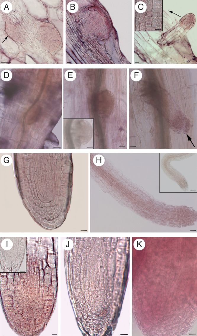
Fig. 7.

Localization of trans-zeatin riboside (A–C) and YUCCA6 transcription (D–H) during AR development in planta [14-day-old Col (A–F) and Ws (G, H) seedlings grown under HF treatment], and YUCCA6 transcription in ARs from TCLs cultured with/without IBA + Kin (I–K). (A) Stage IV (arrow) and stage VI ARPs showing a diffuse cytokinin immunostaining. (B) ARP just before protrusion showing high cytokinin immunostaining in the protoderm, in particular. (C) Elongating ARP with extensive cytokinin signal at the tip. Differentiating epidermis with staining (arrow) magnified in the inset (longitudinal tangential section). (D–E) In situ hybridizations showing YUCCA6 transcription at stage IV (D) and VI (E). Sense probe control shown in the inset of E. (F–H) YUCCA6 transcription at the tip (arrow) of protruded ARPs (F), all over the AR apex (G), and in the AR procambium and differentiating vasculature (H). Sense probe control shown in the inset of H. (I) YUCCA6 transcription in AR apices from IBA + Kin-cultured Ws TCLs. The sense probe control is shown in the inset. (J, K) Low (J) and high (K) YUCCA6 transcription in the apices of ARs formed by sur2-1 TCLs cultured without hormones and with IBA + Kin, respectively. (D–F, H, K) Whole-mount RNA in situ hybridizations; (G, I, J) RNA in situ hybridizations on longitudinal sections of resin-embedded ARs. Scale bars: (inset in C, G, I–K) = 10 µm; (A–C, inset in I) = 20 µm; (D–F, inset in E, H) = 30 µm; (inset in H) = 40 µm.
