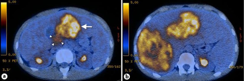
Fig. 1.
a PET/CT showing the primary tumor in the pancreatic head, 3 cm in diameter with weak fluorodeoxyglucose avidity (standard uptake value 3.8; arrowheads). In addition, a large liver metastasis can be seen in the left liver lobe (arrow). b Multiple large liver metastases are shown predominantly in the right liver lobe.

