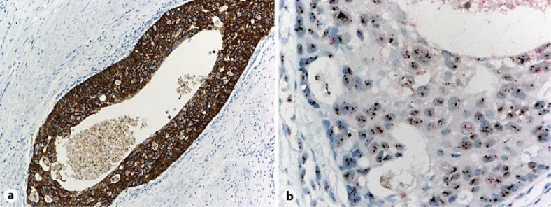
Fig. 2.
a Immunohistochemical analysis revealed that the tumor cells were positive for HER2 overexpression (Dako, Herceptest). b Dual-color chromogenic in situ hybridization revealed that HER2 gene was highly amplified, with HER2:chromosome 17 centromere ratio at 10:1 or more (Ventana, INFORM HER2 Dual ISH). Red = Chromosome 17 centromere; black = HER2.

