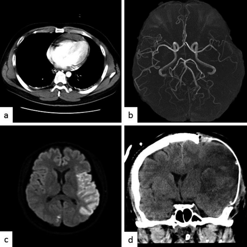
Fig. 1.
a Enhanced chest CT scan on admission shows pericardial effusion. MR angiography shows the occlusion of the superior division of the left M2 segment of the MCA (b) and diffusion-weighted MR imaging shows high signal intensity in the MCA territory (c). d Postoperative CT scan shows diffuse brain edema after the hemispheric infarction; decompressive craniectomy was performed.

