
Fig. 1.
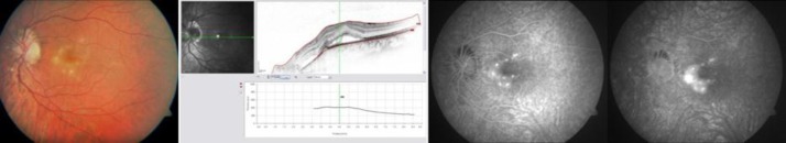
Retinography of the LE showing multiple, round and yellowish lesions in the macula and nasal to the optic nerve (left). OCT with detachment of the neuroepithelium and a slight RPE detachment (middle) and FA revealing hyperfluorescent lesions (right) at presentation.