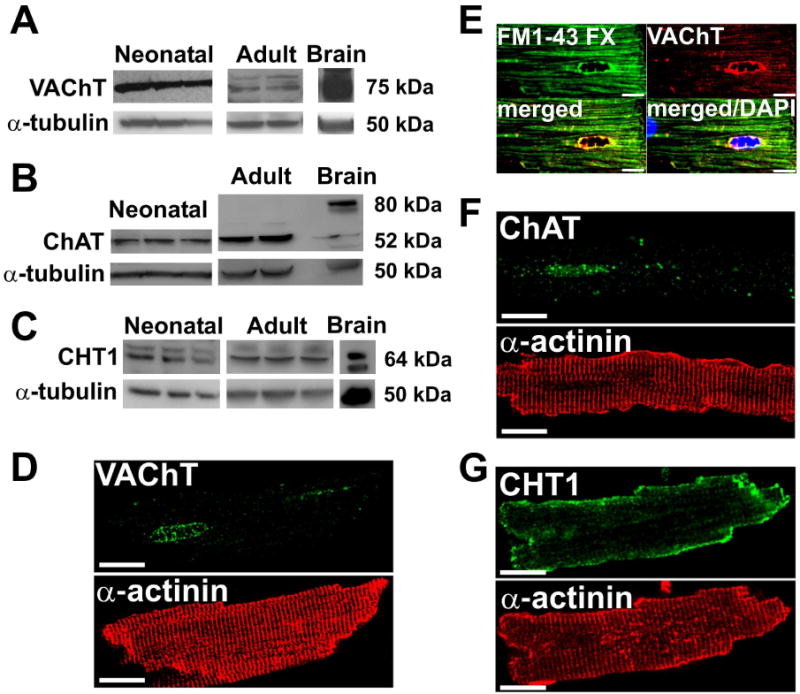
Figure 1. Mouse cardiomyocytes express prototypical cholinergic markers.

A-C. Representative Western blots of immunoreactivity for VAChT, ChAT, and CHT1 in neonatal and adult mouse ventricular myocytes. Brain samples were used as a positive control. α-tubulin was used as a loading control. D-G. VAChT, FM1-43 FX, ChAT and CHT1 staining in adult mouse ventricular myocytes. Cardiomyocytes were also labeled with antibody against the sarcomeric protein α-actinin to show cellular organization. Scale Bar=10 μm.
