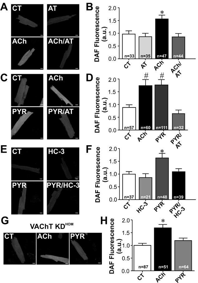
Figure 2. NO levels can be used as a biosensor to detect ACh release in cardiomyocytes.

A-F. DAF fluorescence measurements performed in wild-type (WT) cardiomyocytes. A. Sample confocal images show cellular increase in DAF fluorescence in cardiomyocytes from WT mice treated with ACh. B. Averaged DAF fluorescence increase in adult ventricular myocytes following acute treatment with exogenously added ACh for 30 minutes. The pre-incubation of muscarinic receptor antagonist, atropine (AT), inhibits the increase in DAF fluorescence in ACh treated cardiomyocytes. C. Sample images of DAF fluorescence of cardiomyocytes incubated with ACh, pyridostigmine (PYR) or PYR/AT. D. Effect of cholinesterase inhibition on DAF fluorescence in the presence or absence of atropine (AT) pre-treatment. Cardiomyocytes exposed to pyridostigmine presented an increase in DAF fluorescence, which was blunted by atropine. E. Sample images of DAF loaded cardiac myocytes treated with hemicholinium-3 (HC-3) and/or PYR for 30min. F. HC-3 blunted PYR induced increase in DAF fluorescence in ventricular myocytes. G. Sample images of DAF fluorescence in ventricular myocytes of VAChT KDHOM mice incubated with ACh or PYR. H. Cholinesterase inhibition in cardiomyocytes with reduced VAChT expression levels does not lead to a significant increase in NO generation. n= number of cells analysed. * p < 0.05 when compared to the other groups. # p<0.05 when compared to CT and PYR/AT groups. All cells were loaded with fluorescent dye following the same protocol, and imaging was done preserving the same parameters in both control and drug treated cardiomyocytes. Scale Bar= 10 μm.
