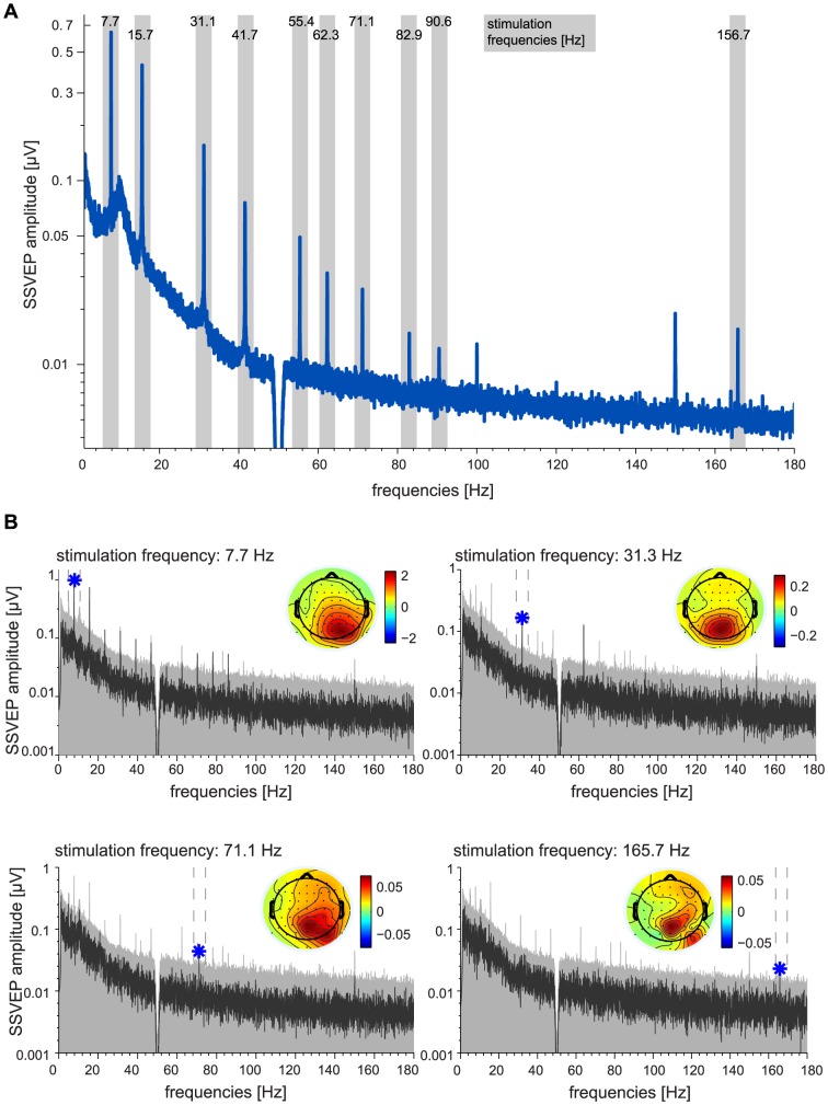
Figure 2. Steady State Visual Evoked Potentials.
A: Grand-average frequency spectra for 30 participants as recorded during phase I, based on 30 s stimulation intervals and averaged across all channels. Gray shades indicate a ±2 Hz range around stimulation frequencies (7.7 Hz to 165.7 Hz). Note that spectral peaks indicating steady state evoked potentials were found even at the highest stimulation frequencies that were never perceived as flickering. B: Data of an exemplary participant. Frequency spectra evoked by four stimulation frequencies: 7.7 Hz, 31.3 Hz, 71.1 Hz, 165.7 Hz (dark gray). The light gray area indicates the 99.9%-percentile of the resampled data, as used to determine statistical significance of peaks at the stimulation frequency. Blue stars indicate significant amplitude peaks at the stimulation frequency (p<0.001). The topographies show the scalp distribution of amplitudes at the peak frequency.

