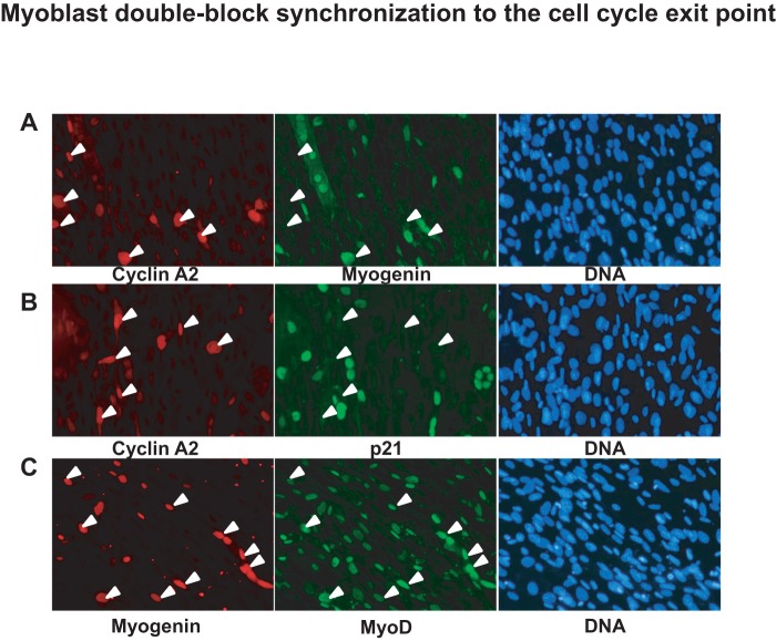
Figure 3. Myoblast double-block synchronization to the cell cycle exit point – Part 2.
C2.7.4 myoblasts were fixed 24-block release. Immunofluorescence was performed revealing either cyclin A2 with myogenin (A), cyclin A2 with p21 (B) and myogenin with MyoD (C). Secondary Alexa-fluor-488 and −455 antibodies were used for immunofluorescence detection and DNA was labeled with Hoechst. White arrows shows cyclin A2 positive cells (A and B) and myogenin positive cells (C).

