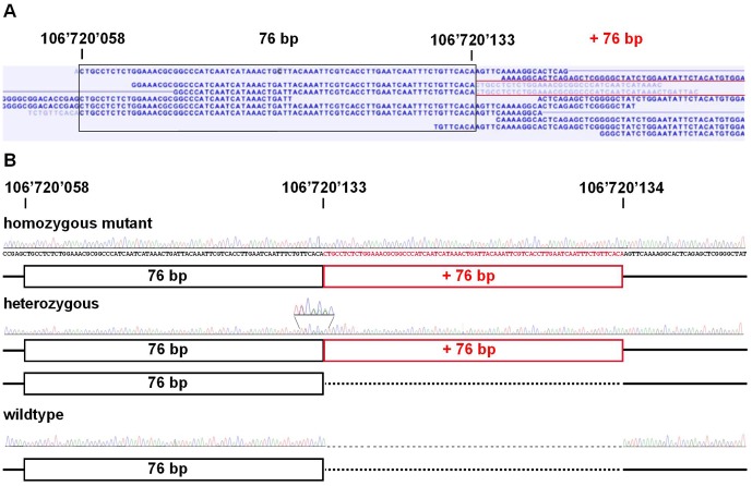
Figure 3. Mutation detection by whole genome re-sequencing.
(A) Screenshot of the short read mapping against the reference sequence, note the red labeled reads indicating the genomic duplication. (B) Sanger sequencing confirmed the presence of the duplication; black box: 76 bp element in the reference sequence; red box: additional 76 bp duplication, note the enlarged region showing the heterozygous sequence using reverse PCR primer for sequencing.

