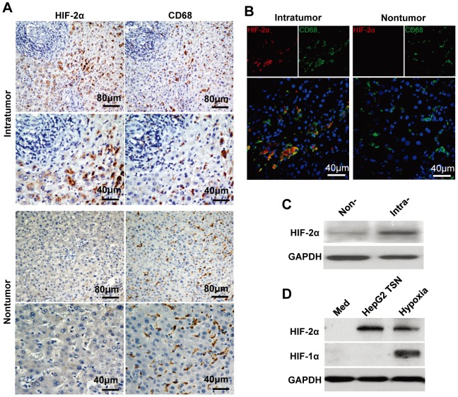
Figure 1. Upregulation of HIF-2α expression in TAMs.
(A) Adjacent sections of paraffin-embedded HCC tissue (n = 9) were stained with an anti-HIF-2α or anti-CD68 antibody. (B) HIF-2α expression in macrophages from frozen sections of HCC tissue (n = 6) was analyzed by confocal microscopy. HIF-2α, red; CD68, green; DAPI, blue. (C) Level of HIF-2α protein in CD14+ cells isolated from non-tumoral (Non-) or intratumoral (Intra-) tissue of 4 HCC patients was analyzed by immunoblotting. (D) Healthy PBMC-derived monocytes were treated with medium alone (Med) for 7 days, or with HepG2 TSN under normoxic condition (HepG2 TSN) for 7 days, or with medium alone for 6 days and then exposed to 1% O2 for another 24 h (hypoxia). Expression of HIF-2α and HIF-1α was determined by Western blotting. This data shown are representative of four separate experiments.

