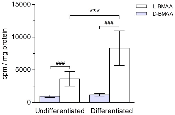
Figure 5. Influx of [14C]-labeled L- and D-BMAA in mammary gland HC11 cells.
Levels of radioactivity in cultured undifferentiated and differentiated mammary gland cells exposed to [14C]-labeled BMAA (1 µM) during 15 min. [14C]L-BMAA is taken up at a significantly higher rate than [14C]D-BMAA in both undifferentiated and differentiated cells. Differentiation of the mammary gland cells to a secretory state significantly increases the uptake of [14C]L- but not [14C]D-BMAA. Mean values (cpm/mg protein) from seven experiments ± SD are plotted. ***p<0.001 compared to undifferentiated cells, and ### p<0.001 compared to D-BMAA within same cell phenotype.

