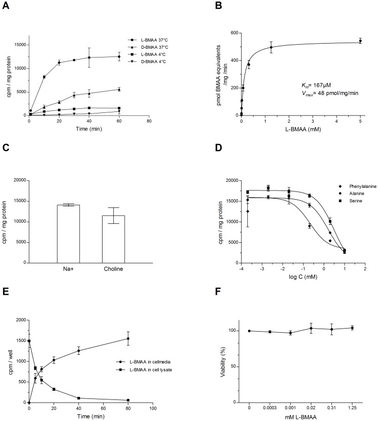
Figure 6. Transport of L- and D-BMAA in differentiated mammary gland HC11 cells.
A Influx of 1 µM [14C]L-BMAA or [14C]D-BMAA up to 60 min exposure in cultured mammary gland cells. The influx was highly reduced at incubation at 4°C, compared to incubation at 37°C. B Concentration dependent influx of [14C]L-BMAA in the presence of unlabeled L-BMAA (0.0003–5 mM) during 15 min exposure in cultured mammary gland cells. The influx of L-BMAA was saturable. C Influx of 1 µM [14C]L-BMAA or [14C]D-BMAA during 15 min exposure in cultured mammary gland cells. Replacement of sodium ions with 120 mM choline chloride did not have a major impact on the influx of [14C]L-BMAA. D Influx of 1 µM [14C]L-BMAA or [14C]D-BMAA during 15 min exposure in cultured mammary gland cells. The presence of competing amino acids reduced the influx of [14C]L-BMAA. E Efflux of 1 µM [14C]L-BMAA up to 80 min from cultured preloaded mammary gland cells. A time-dependent efflux is illustrated by decreasing concentrations in the cells and increasing concentrations in the cell medium. Representative experiments are shown in A-E. F Viability of mammary cells exposed to increasing concentrations of L-BMAA for 3 h, as determined by MTT assay. Data are presented as mean values (% of vehicle) ± SD (n = 3-4/data-point).

