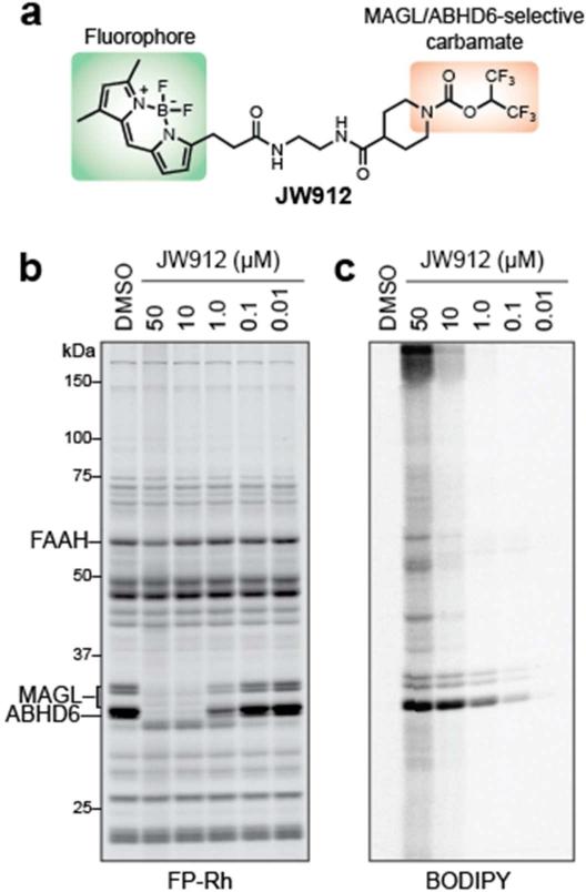
Figure 6.
Development of an activity-based imaging probe for MAGL and ABHD6. (a) Structure of JW912 imaging probe highlighting the HFIP carbamate group, which directs this probe to MAGL and ABHD6, and the BOPIDY fluorophore, which allows visualization. (b) Competitive ABPP for JW912 showing selective inhibition of MAGL and ABHD6 over other serine hydrolases in the brain. (c) BODIPY channel gel image revealing that JW912 selectively labels MAGL and ABHD6 at concentrations below 10 μM across the proteome.

