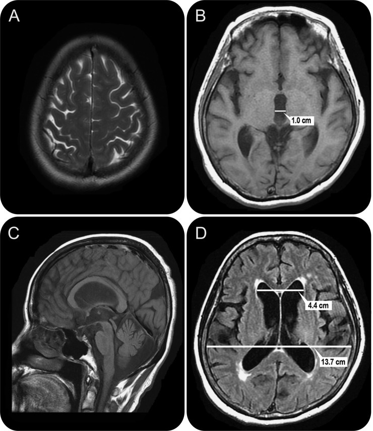
Figure. MRI of a 73-year-old woman with impairment of gait and balance, bladder control, and cognition for 3 years.

The patient had not improved with treatment for parkinsonism. A trial of CSF removal via external lumbar drainage produced substantial gait improvement, and she was treated with a ventriculoperitoneal shunt. At baseline, the Tinetti score was 12–16/28, which is significantly impaired; 9 months after shunt surgery, the score was 26/28, and the patient walked effortlessly. (A) Axial T2 imaging consistent with the Japanese “high and tight” criteria for the convexity. The interhemispheric fissure is effaced. (B) Axial T1 imaging shows a widened IIIrd ventricle with a span of 10 mm. (C) Sagittal T1 imaging shows bowing of the corpus callosum and a pulsation artifact (flow void) in the Sylvian aqueduct. (D) Axial fluid-attenuated inversion recovery imaging shows measurement of the Evans ratio. The diameter of the frontal horns is 4.4 cm, the widest brain diameter is 13.7 cm, and the Evans ratio is 0.32.
