Fig. 3.
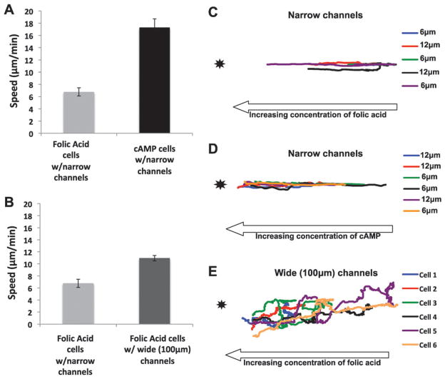
Analysis of cell migration in the OMD. (A) Migration speeds of unpolarized cells within narrow channels are significantly slower than polarized cells within narrow channels toward the chemoattractant source. The migration speed (μm min−1) was measured from a mixture of unpolarized and polarized cells, as described in the materials and methods, toward a micropipette loaded with 10 μM cAMP and folic acid. Ten cells from three different days were observed. The error bars represent the standard error of the mean where n = 30. (B) Unpolarized cells in the presence of narrow channels migrate slower than folic acid cells in the 100 μm wide channels toward folic acid. The migration speed (μm min−1) was measured for the vegetative cells in a device containing narrow channels and a device containing 100 μm wide channels toward 10 μM folic acid. Thirty cells were analyzed, taking ten cells each from three different days. The error bars represent the standard error of the mean where n = 30. (C) Unpolarized cells within the narrow channels migrate in a relatively straight line toward the attractant source. The unpolarized cells were tracked from the videos analyzed in Fig. 3A. Cells from the two innermost 6 and 12 μm channels in the device were analyzed. Five cells were analyzed from three different days. (D) Polarized cells within the narrow channels migrate in a straight line toward the attractant source. The polarized cells were tracked from the same videos analyzed in Fig. 3A. Cells from the two innermost 6 and 12 μm channels in the device were tracked. Six cells were analyzed from three different days. (E) Unpolarized cells in the wide channels migrate in a biased random walk toward the attractant source. The unpolarized cells were tracked from the same videos analyzed in Fig. 3B. Six cells were analyzed from two different days.

