
Figure 2.
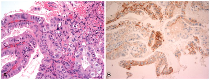
Example of a case with discordance between manual human epidermal growth factor receptor 2 (HER2/neu) immunohistochemistry (IHC) score and HER2/neu image analysis interpretation. A, Biopsy specimen of gastroesophageal junction adenocarcinoma. B, The tumor demonstrates scattered tumor clusters (>5 cells) with HER2/neu IHC 3+ staining, which would be considered overexpression in this biopsy sample. The Automated Cellular Imaging System III (ACIS III) image analysis HER2/neu value was 1.2, which corresponds to a negative interpretation (hematoxylin-eosin, original magnification ×400 [A]; HER2/neu IHC stain [HercepTest, Dako, Carpinteria, California], original magnification × 200 [B]).