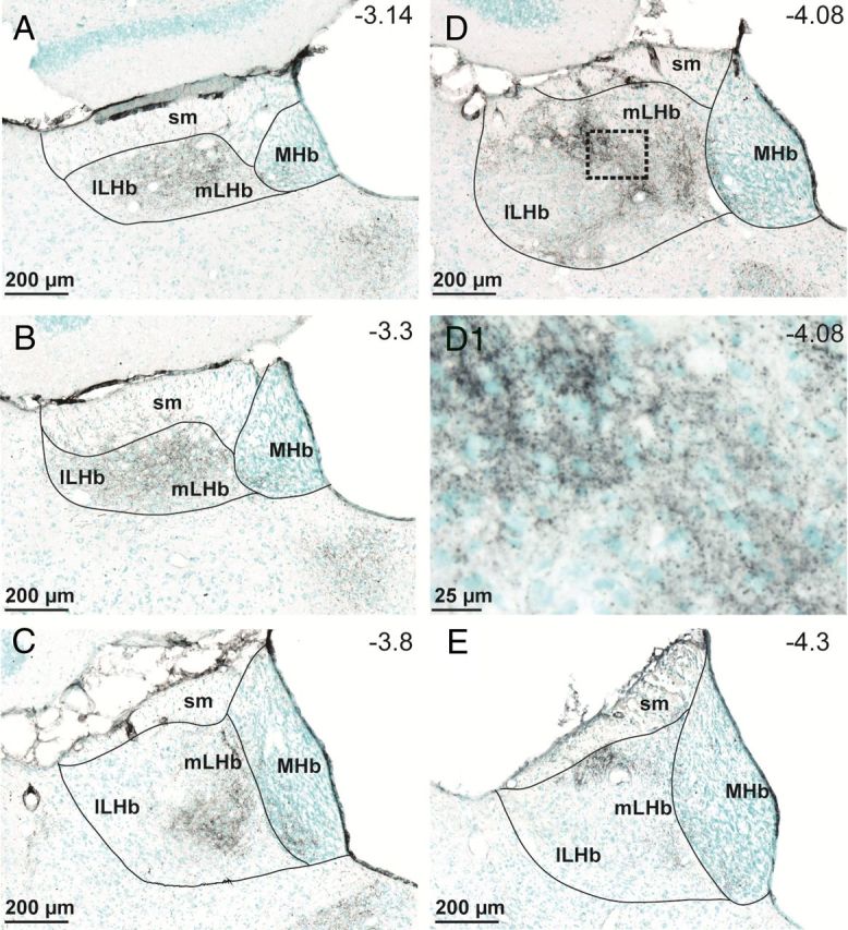
Figure 1.

TH fiber innervation of the LHb. TH-immunopositive fibers are stained in black, whereas cell bodies are labeled with methyl-green. A–E, TH fiber density is highest in an area that we designate the medial lateral habenula (mLHb) (A–E), and our recordings were made in this area as represented in C and D. D1, Higher-resolution image from the area designated by the dashed box in D, showing TH varicosities surrounding LHb neuron somata. Anterior–posterior coordinates from bregma (−3.14 to −4.3 mm) are estimated from the stereotaxic atlas of Paxinos and Watson (1998). sm, Stria medullaris; lLHb, lateral LHb; MHb, medial habenula.
