
Figure 2.
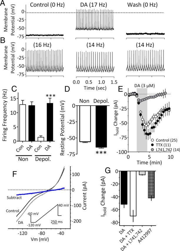
DA depolarizes a population of mLHb neurons. A, Current-clamp recording from an mLHb neuron demonstrating membrane depolarization and an increase in spontaneous action potential frequency caused by bath application of DA (3 μm). B, Absence of DA effect on a mLHb neuron. C, Mean (± SEM) effects of DA (3 μm) on mLHb neuron firing rates. Note that a significant increase in cell firing was observed only in LHb neurons that demonstrated low action potential frequencies before DA application (Depol.). D, Mean RMPs of mLHb neurons that were either depolarized by (Depol.) or insensitive to (Non) DA. Neurons that were depolarized by DA exhibited significantly larger mean RMP. E, Time course of IDAi under control conditions or following application of TTX (500 nm) or the DA D4R antagonist L741,742 (200 nm). The effect of TTX was not significant, whereas that of L741,742 was significantly different from that observed with DA alone. DA application is noted by the vertical shaded bar. F, I–V relationships generated during peak IDAi demonstrate that whole-cell conductance was increased at membrane voltages negative to approximately −45 mV and that IDAi (blue line) had a mean reversal potential of −51.0 mV (95% CI = −50.9 mV to −51.6 mV; linear regression, R2 = 0.99; n = 9 neurons). I–V curves were obtained by slowly changing the membrane potential from −120 to −40 mV using the indicated voltage ramp protocol (inset). The subtracted current was obtained by subtracting the curve obtained during DA from that obtained during the control condition. G, The effects of DA (3 μm) on Ihold, alone, or in the presence of TTX (500 nm) or L741,742 (200 nm) compared with those of the selective D4R agonist A412997 (1 μm; n = 6). DA alone, and in TTX, and A412997 significantly increased Ihold (F(3,45) = 16.44, p < 0.0001, one-way ANOVA). However, DA did not significantly affect Ihold after L741,742 treatment.