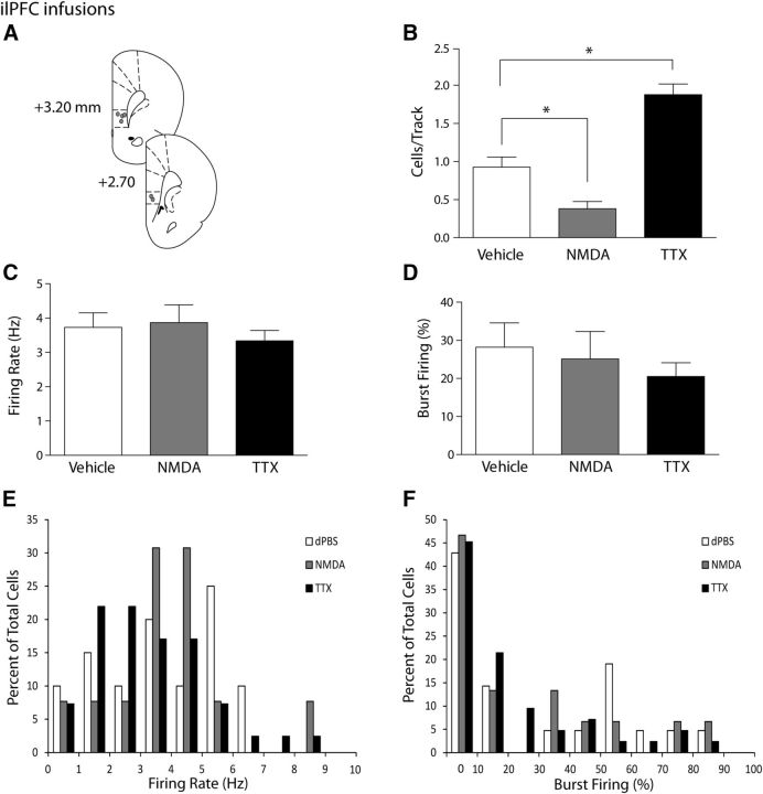
Figure 2.
Activation or inactivation of the ilPFC selectively decreases or increases VTA DA neuron population activity, respectively. A, Representation of histological placements of infusion cannulae in the ilPFC (circles, ∼50% shown). B, Activating the ilPFC with NMDA resulted in a significant decrease in the number of spontaneously active DA neurons in the VTA (cells/track, gray bar), whereas inactivating the ilPFC with TTX yielded a significant increase in the number of spontaneously active DA neurons in the VTA (black bar), compared with vehicle injections (white bar). C, D, Infusion of vehicle (white bars), NMDA (gray bars), or TTX (black bars) led to no changes in the firing rate or percentage of spikes in bursts in dopaminergic cells in the VTA. Distributions of firing rate (E) and the percentage of burst firing (F) were not affected significantly by infusions of NMDA or TTX (Kolgomorov–Smirnoff test). *p < 0.05 (one-way ANOVA, Holm-Sidak post hoc test). n = 4 or 5 rats/group; n = 15–42 neurons/group. Data are represented as mean ± SEM.

