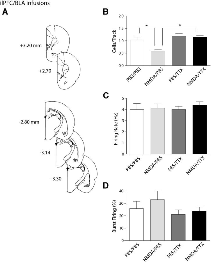
Figure 3.
The ilPFC activation-induced decrease in VTA DA neuron population activity is mediated via the BLA. A, Histological placement of infusion cannulae into the ilPFC and BLA (circles, ∼50% of ilPFC placements [top] shown for clarity and 100% of BLA placements [bottom] shown). B, Dual infusions of NMDA into the ilPFC and TTX into the BLA (black bar) prevented the decrease in number of spontaneously active dopamine cells in the VTA that occurred with activation of the ilPFC alone (cells/track, light gray bar). Inactivating the BLA alone did not yield any changes in dopamine population activity compared with vehicle infusions (dark gray bar and white bar, respectively). C, D, The firing rate and percentage of spikes occurring in bursts were not affected by any of the infusions. *p < 0.05 (two-way ANOVA, Bonferroni post hoc test). n = 5 rats/group; n = 18–41 neurons/group. Data are represented as mean ± SEM.

