
Figure 1.
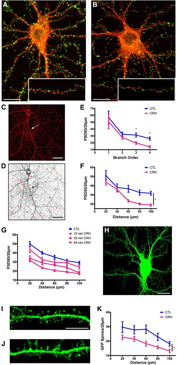
The stress neuropeptide CRH causes a loss of PSD95-ir puncta and dendritic spines in cultured rat hippocampal neurons. Exposure to 100 nm CRH leads to a significant reduction in PSD95-ir puncta, an indication of dendritic spine loss. A, B, Control neuron and (B) neuron exposed to 100 nm CRH at 36°C for 30 min and processed for ICC for PSD95 (green) and for F-actin (red). C, Confocal images were used to quantify PSD95-ir puncta and dendritic spines from GFP-expressing neurons. Neurons used for quantification were clearly demarcated and devoid of dendritic crossings from other neurons that could confound counts. D, Example of a confocal image processed for quantification with 20 μm segments measured out from the soma. E, Exposure to CRH reduced the density of PSD95-ir puncta along dendritic branches (F(1,66) = 3.81, p = 0.006), and this effect became more apparent in third-order (p = 0.004) and fourth-order (p < 0.001) branches (n = 12). Similar results were obtained by quantifying PSD95 by distance from the soma. F, Graph quantifying PSD95-ir puncta per 20 μm segment in cultures incubated in the presence or absence of CRH (F(1,88) = 7.09, p = 0.014; n = 12). G, CRH reduced PSD95 puncta in a time-dependent manner (F(3,80) = 17.66, p < 0.001; n = 6). H, Lentiviral infection of neurons did not change the size or shape of the soma, and enabled direct visualization of spines. I, J, An example of a control GFP-filled dendrite and (J) a dendrite after exposure to 100 nm CRH at 36°C for 60 min. K, Graph quantifying GFP-filled spines per 20 μm segment with and without exposure to CRH (F(1,48) = 7.55, p = 0.017; n = 12). The values for spine density were approximately half of those found using PSD95-ir puncta because the GFP analysis is limited to spines perpendicular to the dendrite. Scale bars: A–D, H, 20 μm; I, J, 5 μm.