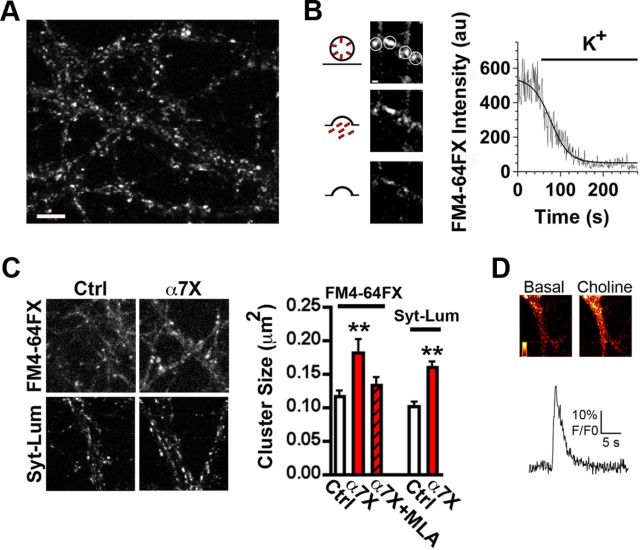
Figure 3.
Reducing the mobility of α7–nAChRs increases the pool size of recycling vesicles. A, Representative image of FM4-64FX staining in hippocampal cultures. Scale bar, 5 μm. B, Left, Representative image of a presynaptic terminal at various times after stimulation with KCl. Right, Trace showing a representative destaining time course following a sigmoidal kinetic (solid line) in arbitrary units (au). C, Left, Representative images of FM4-64FX and Syn–Lum staining in control (Ctrl) and crosslinked (α7X) conditions. Right, Quantification of FM4-64FX cluster size in Ctrl, α7X, and α7X plus MLA, or Syt–Lum cluster size in Ctrl and α7X (n = 21–24 neurons from 4–5 culture sets for each condition; mean ± SEM; one-way ANOVA, **p < 0.01). D, Top, Representative fluorescent images of a dendrite loaded with the calcium fluor Fluo-4 captured before (Basal) and after (Choline) puffing on choline. Bottom trace shows a representative time course for the decay of the fluorescent signal after α7–nAChR activation by choline.

