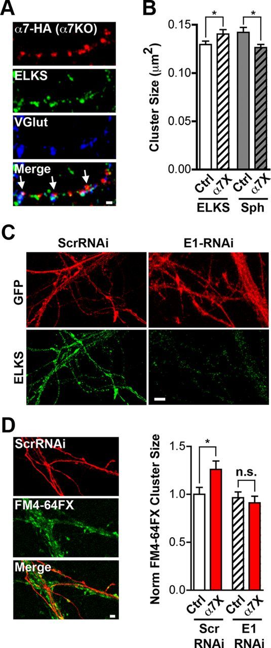
Figure 8.

Functional proteomics suggests ELKS as a possible mediator of α7–nAChR crosslinking effects on release capacity. A, Representative images of an axon from a transfected neuron expressing HA–α7–nAChR (red), ELKS (green), and VGluT immunostaining (blue) and the three merged (Merge) with arrows indicating colocalization characteristic of presynaptic terminals. Scale bar, 1 μm. B, Quantification of ELKS and synaptophysin (Sph) cluster size on rat hippocampal neurons in culture under control (Ctrl) and crosslinked α7–nAChR (α7X) conditions (n = 27 neurons, 4 culture sets per condition; mean ± SEM; Student's t test, *p < 0.05). C, Representative images of neurons expressing GFP (red) and ELKS (green) after being infected with either control Scr–RNAi or E1–RNAi virus. Scale bar, 5 μm. D, Left, Representative images of cells expressing control Scr–RNAi (red), stained for FM4-64FX (green), and the images merged (Merge). Scale bar, 2 μm. Right, Quantification of FM4-64FX puncta size in Ctrl and α7X cultures infected with either Scr–RNAi virus as control or E1–RNAi virus (n = 25 neurons, 4 culture sets per condition; mean ± SEM; Student's t test, *p < 0.05).
