
Figure 2.
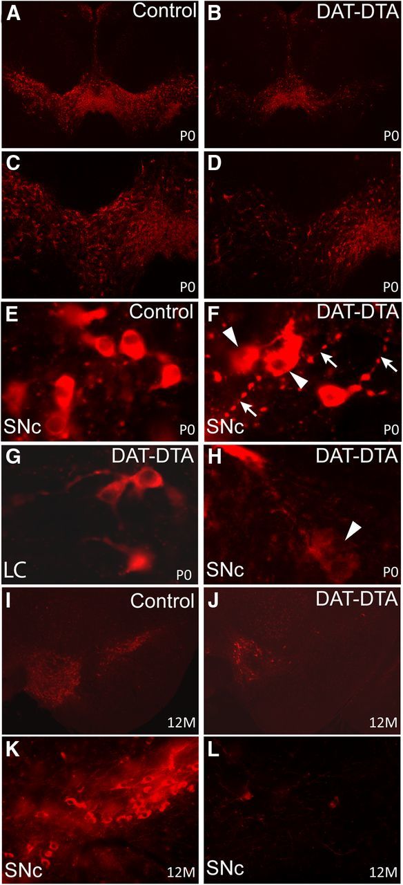
Loss of TH-positive neurons in the ventral midbrain of DAT-DTA mice. A–D, Fewer TH-positive neurons are observed in the SNc and VTA of DAT-DTA mice at P0 compared with control mice. TH-positive neuron loss is less severe in the VTA at this age. E, F, H, At P0, TH-positive neurons in the SNc of DAT-DTA mice (F, H) appear abnormal compared with neurons in the SNc of control mice (E). TH-positive neurons in the SNc of DAT-DTA mice (F, H) appear to be actively degenerating (arrowheads) and have neurites with large varicose swellings (F, arrows). G, TH-positive neurons in the locus ceruleus (LC) of DAT-DTA mice have a normal appearance. I–L, At 12 months of age, fewer TH-positive neurons are observed in the SNc and VTA of DAT-DTA mice compared with control mice. TH-positive neuron loss appears to be more severe at 12 months compared with P0 in both the SNc and VTA of DAT-DTA mice.