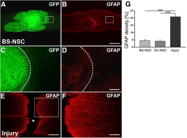
Figure 3.
Grafted BS-NSCs survive and integrate in the host parenchyma, reducing injury-induced astrogliosis. A–D, The lesion site at level T4 is completely filled with GFP-labeled implanted BS-NSCs. Grafted cells integrate in the rostral and caudal host spinal cord. GFAP expression is low in BS-NSC grafts and the surrounding host spinal cord. At higher magnification (D), weak GFAP labeling can be observed at the host/graft interface identified by GFP immunolabeling (C, dotted lines). E, F, In the spinal cord of animals that received no graft, a lack of GFAP labeling in the lesion site (asterisk) and an upregulation of GFAP in spinal parenchyma adjacent to the lesion site representing the glial scar is visible. Boxed regions in A, B, and E are shown at higher magnification in C, D, and F, respectively. G, Compared with animals that received an injury but no graft, the density of GFAP immunolabeling at the lesion border is significantly lower in animals grafted with BS-NSCs or SC-NSCs (ANOVA, p < 0.0001 with Fisher's PLSD, ***p < 0.0001). Scale bars: A, B, 1 mm; C, D, F, 200 μm; E, 0.5 mm.

