Figure 8.

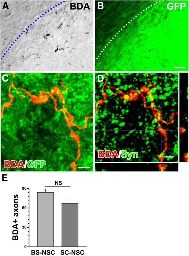
Host supraspinal projections connect with grafted BS-NSCs in the spinal cord. A, B, BDA-labeled rostroventrolateral medulla-derived vasomotor axons (black) regenerate into the graft identified by GFP immunolabeling (B, green). The host spinal cord/BS-NSC graft interface is indicated by a dotted line. C, Confocal fluorescent microscopy demonstrates terminals of BDA-labeled descending projections (red) within BS-NSC implants, displaying bouton-like structures. D, Confocal analysis reveals that the terminals of BDA-labeled fibers (red) in the graft colocalize (yellow) with the presynaptic marker synaptophysin (green), suggesting the formation of functional synapses. E, Quantitative analysis shows that the number of BDA-labeled axons extending into BS-NSC grafts is not significantly (NS) different from those in SC-NSC grafts (unpaired t test, p > 0.05). Scale bars: A, B, 100 μm; C, D, 5 μm.
