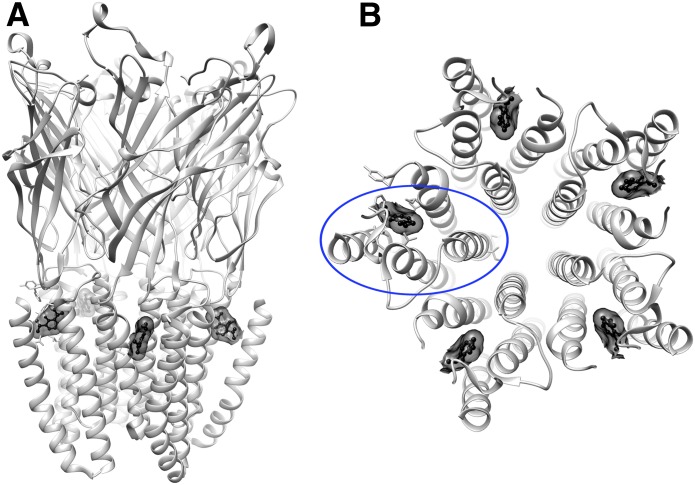
Fig. 1.
GLIC propofol structure (PDB ID 3P50) showing propofol in ball-and-stick and transparent surface representation (black), bound near the top of the helical TM domains within each subunit. (A) Side view. (B) View from the top of the TM domains, with one of the subunits circled in blue.

