
Figure 2.

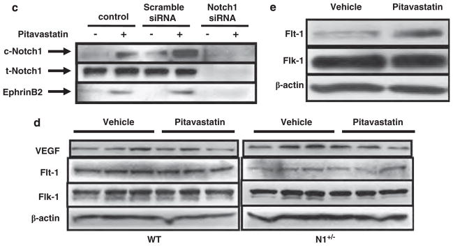
Impaired post-natal arteriogenesis in Notch1 heterozygous-deficient mice (N1 +/−) mice treated with pitavastatin. (a) Top: representative pictures of immunostaining for α-smooth muscle actin (SMA) and CD31 for new vessels from eight individual mice from each group. Bar = 10 μm. Bottom: arterial perimeter and diameter quantitative data in ischemic limbs from wild-type (WT) and N1 +/− mice with the vehicle and pitavastatin treatment and control limbs. *P<0.05 compared WT mice with the vehicle treatment. **P<0.05 compared WT mice with the pitavastatin treatment. (b) Left: representative photomicrographs of αSMA-positive arterioles in the ischemic adductor muscles of WT and N1 +/− mice, with the vehicle or pitavastatin treatment from eight individual mice from each group. Bar = 50 μm. Right: quantitative analysis of arteriole density (five microscopic fields from four different sections per mice; eight mice each; number/h.p.f.; ×200 magnification). *P<0.05, compared with WT with the vehicle treatment. **P<0.001 compared with WT mice with the pitavastatin treatment. (c) Immunoblots showing total and cleaved Notch1 (t- and c-Notch1), and Ephrin-B2 expression in human umbilical vein endothelial cells (HUVECs) transfected with scramble or Notch1 small-interfering RNA (siRNA) in the absence or presence of pitavastatin (100 nmol/l, 2 h). Representative data of three independent experiments with similar results. (d) Immunoblots showing VEGF, Flt-1 and Flk-1 expression in ischemic limbs from WT and N1 +/− mice in the absence or presence of pitavastatin at 3 post-operative days. Representative data of three independent experiments with similar results. (e) Immunoblots showing Flt-1 and Flk-1 expression in HUVECs in the absence or presence of pitavastatin. Representative data of three independent experiments with similar results.