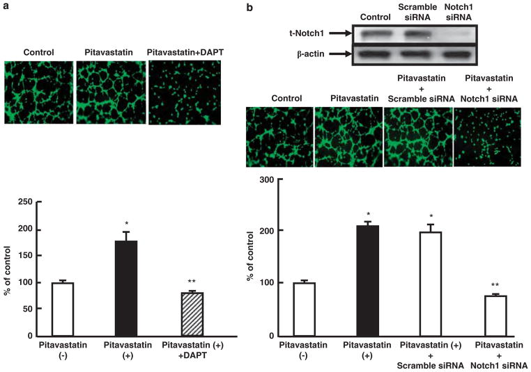
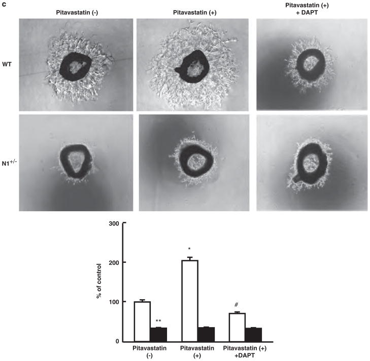
Figure 7.
Notch1 mediates tube formation and microvascular sprouting in vitro. (a) Top: representative photomicrographs (× 400 magnification) showing Matrigel-based, capillary-like tube formation in human umbilical vein endothelial cells (HUVECs) treated with pitavastatin (100 nmol/l) and γ-secretase inhibitor DAPT (20 μmol/l). Bottom: quantitative data of capillary-like tube formation. Data shown are mean ± s.d., n = 5 in each group. *P<0.01 compared with the absence of pitavastatin and **P<0.01 compared with pitavastatin only. (b) Top: immunoblots showing the expression of Notch1 and β-actin in HUVECs transfected with scramble or Notch1 small-interfering RNA (siRNA). Center: representative photomicrographs (× 400 magnification) showing Matrigel-based, capillary-like tube formation in HUVECs transfected with scramble or Notch1 siRNA in the presence of pitavastatin (100 nmol/l). Bottom: quantitative data of capillary-like tube formation. Data shown are mean ± s.d., n = 5 in each group. *P<0.01 compared with the absence of pitavastatin and **P<0.01 compared with pitavastatin plus scramble siRNA. (c) Top: representative fields (× 40 magnification) showing microvessel outgrowth in response to pitavastatin (100 nmol/l) and DAPT (20 μmol/l) in aortic explants from wild-type (WT) and Notch1 heterozygous-deficient mice (N1+/−) mice. Bottom: microvessel outgrowth was quantified as percentage of that in WT in the absence of pitavastatin. Data shown are mean ± s.d., n = 5 in each group, *P<0.001 compared with WT in the absence of pitavastatin ,**P<0.001 compared with WT in the absence of pitavastatin and #P<0.001 compared with WT with pitavastatin.


