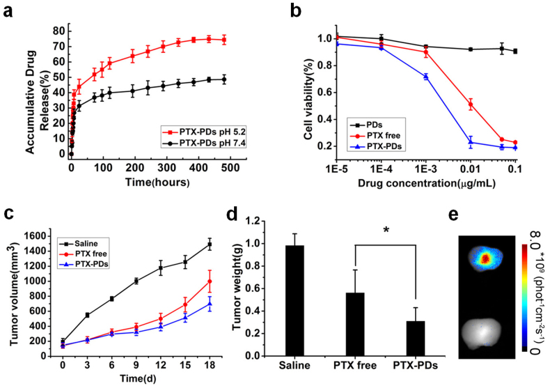
Figure 5.
(a) In vitro drug release of PTX-loaded PDs in acidic conditions (pH 5.2) and physiological conditions (pH 7.4). (b) Cytotoxicity of free PTX, PTX-loaded PDs, and blank PDs after 48 h incubation with MCF-7 cells. The concentration of blank PDs used was equal to the concentration of PTX-loaded PDs. (c, d) Tumor volume and tumor weight of MCF-7 tumor-bearing female nude mice treated with saline, PTX or PTX-loaded PDs. *,P<0.05.(e) Fluorescence images showing the ex-vivo biodistribution of PDs in tumors isolated from the mice after intravenous injection. Lower, saline group; Upper, PTX-loaded PDs group.

