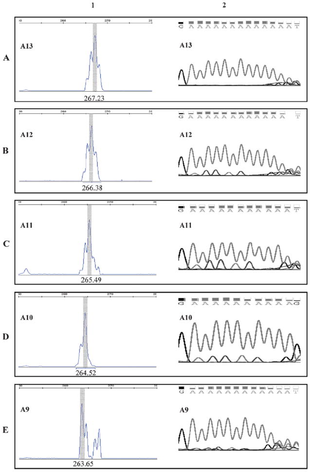
Figure 1.

Deletion of polyA tract in EGFR 3′ untranslated region (3′ UTR) determined by capillary electrophoreses (CE) and Sanger sequencing. The length of EGFR 3′ UTR polyA was measured by CE and confirmed by Sanger sequencing as described in “Materials and Methods.” The size of the polymerase chain reaction fragment with wild-type (WT) 3′ UTR polyA is 267 bases. Column 1, fragment analysis by CE. Column 2, chromatograms of the 3′ UTR polyA regions by Sanger sequencing. A, A13: WT polyA tract with 13 nucleotides. B, A1: polyA tract with 1 nucleotide deletion. C, A11: polyA tract with 2-nucleotide deletion. D, A10: polyA tract with 3-nucleotide deletion. E, A9: polyA tract with 4-nucleotide deletion.
