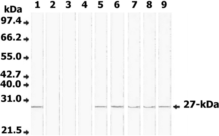
Fig 2.

Western blot of serum samples from Fasciola-infected and noninfected individuals for the detection of circulating Fasciola antigen. Lane 1, FWAP; lanes 2 to 4, serum samples from 3 noninfected individuals; lanes 5 to 9, serum samples from 5 individuals infected with F. gigantica. Fifty micrograms/lane of each sample was separated on 12% acrylamide gels, transferred to an NC sheet, and reacted with 100 μg/ml rabbit anti-27-kDa IgG antibody. Anti-rabbit IgG-alkaline phosphatase and BCIP (5-bromo-4-chloro-3-indolylphosphate)-nitroblue tetrazolium (NBT) substrate were used to visualize the reaction products. The developed rabbit antibody identified a 27-kDa antigen in all sera of infected individuals (lanes 5 to 9) but not in sera of noninfected individuals (lanes 2 to 4). Molecular mass standards included were phosphorylase B (97.4 kDa), bovine serum albumin (66.2 kDa), glutamate dehydrogenase (55 kDa), ovalbumin (42.7 kDa), aldolase (40 kDa), carbonic anhydrase (31 kDa), and soybean trypsin inhibitor (21.5 kDa).
