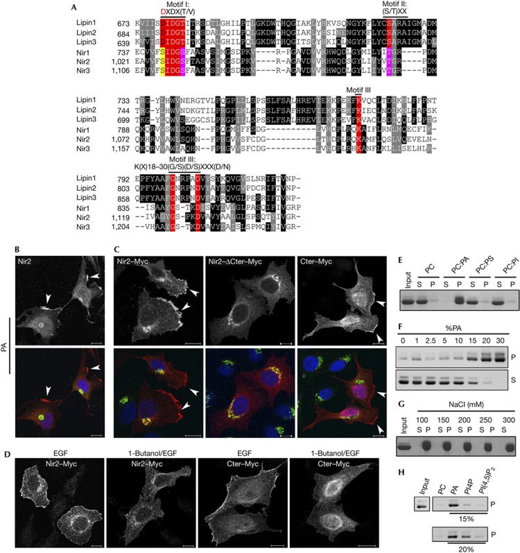
Figure 2.
Phosphatidic acid (PA) triggers the translocation of Nir2 to the plasma membrane (PM) through the C-terminal region that binds to PA. (A) Alignment of the LNS2 (Lipin/Nde1/Smp2) domains of the human lipin and Nir proteins (accession numbers are given in supplementary information online). The alignment was obtained by Clustal W. Black and grey backgrounds represent degree of similarity (black; identity: 14.4%, grey; high similarity: 20.92%), numbers represent aa residues. The three conserved motifs in the haloacid dehalogenase superfamily: Motif I, DXDX(T/V), contains a conserved aspartate residue (red), which acts as nucleophile to form an acylphosphate intermediate in the proposed reaction mechanism for phosphotransferases [20, 21]. In Motif II, S/TXX, the conserved Ser/Thr is involved in hydrogen bonding to the phosphoryl oxygen, whereas Motif III, K(X)18-30(G/S)(D/S)XXX(D/N), is involved in phosphoryl oxygen hydrogen bonding and coordination of the magnesium ion [20]. (B,C) Serum-starved HeLa cells expressing endogenous Nir2 (B) or the indicated Myc-tagged Nir2 proteins (C) were stimulated with PA (100 μM) for 30 min. The cells were then fixed and double immunostained with anti-Nir2 (B) or anti-Myc (C) antibodies (red) together with anti-p115 antibody (green). The localization of endogenous Nir2 (B) as well as Nir2–Myc and its mutants (C) is shown. PM localization is marked by arrowheads. (D) Effect of 1-butanol on the translocation of Nir2. Serum-starved HeLa cells were preincubated with 0.3% 1-butanol for 30 min, and then stimulated with EGF (100 ng/ml) for 10 min in the presence of 1-butanol. The cells were fixed, immunostained with anti-Myc antibody and analysed by confocal microscope. Scale bar, 10 μm. (E) The C-terminal region of Nir2 binds to PA in vitro. The binding of recombinant C-terminal region of Nir2 to multilamellar vesicles consisting of PC alone, PC:PA (2:1), PC:PS (2:1) or PC:PImix (5:1) was assessed by liposome sedimentation assay (Methods). P and S indicate pellet and supernatant fraction, respectively. (F–H) Binding of recombinant C-terminal region of Nir2 to multilamellar vesicles containing either the indicated percentage of PA (F), 33% PA in the presence of the increasing concentrations of NaCl (G), or to vesicles containing 15% or 20% of PA, PI4P or PI(4,5)P2 (H) was examined as described in E.

