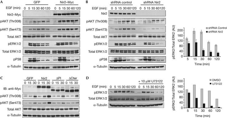
Figure 3.
Nir2 modulates specific signalling pathways downstream to EGFR. HeLa cells were infected with lentiviruses expressing GFP, the Myc-tagged Nir2 (A), control shRNA or Nir2 shRNA (#1) (B). The cells were serum starved for 18 h and then stimulated with EGF (100 ng/ml) for the indicated time periods. Total cell lysates were analysed for phosphorylation of the indicated proteins by western blotting (WB) using the corresponding antibodies. Reproducible results were obtained in at least four experiments. (C) HeLa cells were infected with lentiviruses encoding GFP, Myc-tagged WT Nir2, or its ΔPI or ΔCter mutants. Three days later, the cells were serum starved for 18 h and then stimulated with EGF for the indicated time periods, lysed and immunoblotted with antibodies against the indicated proteins. Shown are representative results of at least four experiments. (D) Serum-starved HeLa cells were pretreated with the PLCγ inhibitor U73122 (10 μM, 30 min), and then stimulated with EGF for the indicated time periods. Total cell lysates were examined for ERK1/2 phosphorylation by WB using anti-pERK1/2 antibody. A.U., arbitrary unit; DMSO, dimethyl sulphoxide; EGF, epidermal growth factor; GFP, green fluorescent protein; IB, immunoblot; PITD, phosphatidylinositol-transfer domain; PM, plasma membrane; shRNA, short hairpin RNA.

