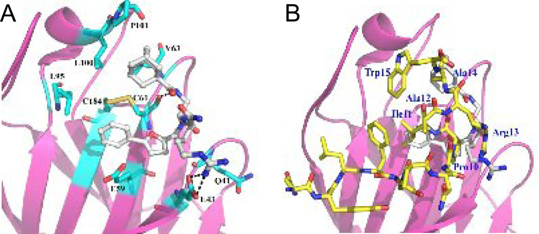
Figure 3.
Molecular modeling of compound 5 in the EphB4 ephrin-binding pocket. (A) The EphB4 receptor is shown as a pink ribbon with residues important for TNYL-RAW binding indicated in cyan and labeled. Compound 5 is shown as gray sticks, and forms hydrogen bonds with EphB4 E43 and C61. Yellow indicates the disulfide bond between EphB4 C61 and C184. (B) Superposition of compound 5 with the TNYL-RAW peptide from the crystal structure of the complex with EphB4 (PDB entry 2BBA). Compound 5 is shown in gray sticks and TNYL-RAW in yellow sticks.

