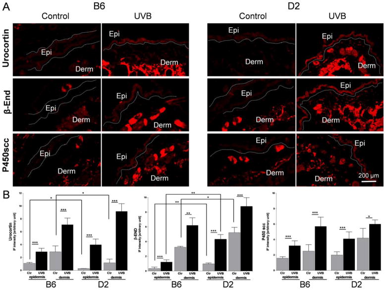
Fig. 3.
Panel A The immunohistochemical detection (immunofluorescent CY3 red fluorophore) of urocortin, β-endorphin (β-END) and P450 cholesterol side-chain cleavage enzyme (P450scc; CYP11A1) performed on cryosectioned skin 24 h after UVB radiation (400 mJ/cm2) with corresponding controls (for antibodies characteristics please see Table 3).
All antigens under investigation were higher expressed after UVB treatment compared to control (sham-treated) scraps. The distribution pattern of immunopositive signals was similar between B6 and D2. Ctr=Control, UVB=UVB irradiated skin, Epi=epidermis, Derm=dermis.
Panel B. Comparison of immunofluorescent signal (IF) intensity for Urocortin, β-END and P450scc between B6 and D2 measured separately in epidermis and dermis with Image J, NIH. 6 measurements per tissue area and 6 biopsies per antigen were investigated and the average results compared between conditions and strains are presented on the graphs. The average IF signal intensities were higher after UVB exposure compared to sham treated (Ctr) samples for all antigens under investigation. The constitutive Urocortin expression was slightly lower in D2 compared B6 while β-End IF signal levels were higher in D2 than B6. The statistical differences were measured with t-test and marked with *, where p<0.05 (*), p<0.01 (**), and p<0.001 (***).

