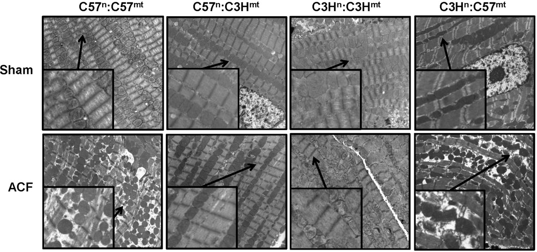
Figure 5. Transmission Electron Microscopy (TEM) of left ventricular (LV) tissue in sham and aortacaval fistula (ACF) treated mice.
Representative images from control (C57n:C57mt and C3Hn:C3Hmt) and MNX (C57n:C3Hmt, and C3Hn:C57mt) mice that underwent either sham or ACF surgery and sacrificed 3 days thereafter, and LV tissue prepared for TEM. Mitochondrial swelling and disorganization along with myofibrillar degeneration was observed in the ACF treated mice having the C57 mtDNA (C57n:C57mt and C3Hn:C57mt animals), which was absent in the ACF treated mice having the C3H mtDNA (C3Hn:C3Hmt and C57n:C3Hmt mice). Mag = 4500x (insets are 9000x, arrows indicate enlarged area). Imaging by Emlabs, Inc., Birmingham, AL.

