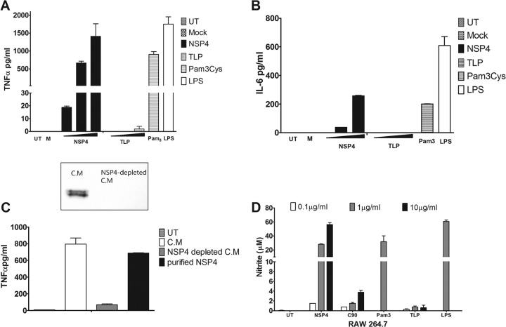
Fig 1.
NSP4 induces secretion of proinflammatory cytokines from THP-1 cells. (A and B) Dose-dependent secretion of TNF-α and IL-6 from pTHP-1 cells. PMA-treated THP-1 cells (2 × 10⁵ cells/well) were incubated with increasing concentrations of purified NSP4 (0.1, 1.0, and 10 μg/ml) or CsCl-purified TLPs (MOI of 1, 10, and 100). The amount of cytokine secreted was compared to that of untreated pTHP-1 cells (UT), cells incubated with medium from uninfected Caco-2 cells (M), or cells treated with control agonists LPS (0.1 μg/ml) and Pam3CSK4 (1 μg/ml) for 24 h. (C) Secretion of TNF-α from pTHP-1 cells in response to 10×-concentrated medium (C.M) from rotavirus-infected cells after ultracentrifugation to remove virus particles and the same medium following immunodepletion of NSP4. Western blot analysis (inset) confirms removal of NSP4 from the concentrated medium. Purified NSP4 (1 μg/ml) was used as a positive control. Cytokine concentrations in the medium were determined by cytometric bead array assay. Data are the means ± SEM of duplicate samples. (D) RAW 264.7 cells were stimulated with increasing concentrations of purified NSP4 (0.1, 1.0, and 10 μg/ml) or identical amounts of CsCl-purified TLPs. Pam3CSK4 (10 μg/ml), LPS (1 μg/ml), and medium from mock-infected Caco-2 cells were used as controls. After 24 h of stimulation, cell-free medium was collected and assayed for nitrite using the Griess assay. Data are means ± SEM from triplicate samples.

