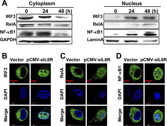
Fig 7.

Effects of sIL6R on IRF3 and NF-κB translocation. (A) A549 cells were transfected with pCMV-sIL6R or a control vector. Then cytosolic and nuclear extracts were prepared at the indicated time points and were subjected to Western blot analysis. GAPDH and lamin A were used as markers for the cytosolic and nuclear fractions, respectively. Levels of IRF3, RelA, and NF-κB1 proteins were detected by Western blotting. (B to D) A549 cells were transfected with pCMV-sIL6R or a control vector for 48 h. After fixation, the cells were immunostained with antibodies against IRF3 (B), RelA (C), and NF-κB1 (D). Nuclei were stained with 4′,6-diamidino-2-phenylindole (DAPI) (blue). Bars, 10 μm.
