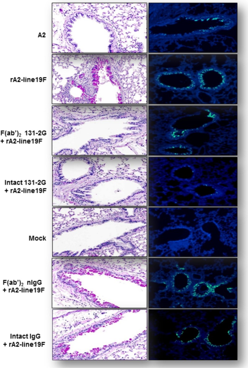
Fig 6.
Mucin production and RSV replication associated with RSV rA2-line19F challenge and anti-G protein MAb 131-2G prophylaxis. BALB/c mice were challenged with mock-infected tissue culture supernatant (mock) or 1 × 10⁶ TCID50 of A2 or rA2-line19F and were untreated or treated 2 days before challenge with the intact or F(ab′)2 form of both intact 131-2G and control immune globulin (nIgG) (n = 5 mice/group). Lungs were harvested 5 days p.i. for RSV antigen immunofluorescence and 8 days p.i. for PAS staining. (A) Representative photomicrographs of PAS-stained lung tissue collected at 8 days p.i. Reddish-purple color (e.g., evident in rA2-line19F-labeled panel) indicates PAS-positive cells indicative of mucin (n = 3 mice/group). (B) Representative photomicrographs of immunofluorescence staining for RSV from the groups of mice noted above at day 5 p.i. Green indicates RSV proteins in the cells (e.g., evident in panels labeled A2 and rA2-line19F; n = 3 mice/group). Results are representative of two independent experiments for untreated or 131-2G F(ab′)2-treated mice challenged with rA2-line19F.

