Fig 3.
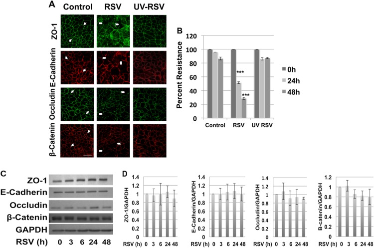
RSV-induced AJC disassembly requires live replicating virus. Polarized 16HBE140- monolayers were infected with RSV at an MOI of 0.5, control medium, or an equal amount of UV-irradiated RSV for 48 h. (A) Apical junction complexes were visualized by immunofluorescent staining and confocal microscopy. The arrows indicate normal chicken wire appearance of membrane AJC. The arrowheads indicate gaps in AJC integrity and ZO-1 localized to intracellular vacuoles. (B) TEER was measured using a volt-ohm meter at the indicated time points. UV-inactivated RSV did not cause epithelial junction disassembly or changes in TEER. The images are representative of at least 3 independent experiments. Differences between control, UV-irradiated RSV-, and RSV-infected cells were analyzed by ANOVA. ***, P < 0.001. The error bars indicate SEM. (C) RSV infection did not change AJC protein expression. Total cell lysates of control and RSV-infected epithelial cells were analyzed by Western blotting with antibodies directed against different TJ and AJ components. Immunoblots representative of different time points postinfection are shown. (D) Densitometric quantification of at least 3 independent experiments.

