
Fig 6.
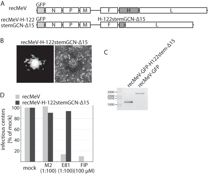
Recombinant MeV expressing the truncated H-122stemGCN-Δ15 attachment protein. (A) Schematic of the standard recMeV and recMeV-H-122stemGCN-Δ15 genomes. Both cDNA constructs harbor an additional eGFP open reading frame in the primary position (light gray). (B) Assessment of cytopathicity induced by recovered recMeV-H-122stemGCN-Δ15 particles 24-h postinfection, using fluorescence and standard microscopy. (C) RT-PCR analysis of the attachment protein-encoding open reading frames after infection of cells with recMeV or recMeV-H-122stemGCN-Δ15. Numbers specify molecular weight of a DNA standard (in bases). (D) recMeV-H-122stemGCN-Δ15 particles are resistant to neutralizing antibodies directed against the H head domain. Virus neutralization assays were conducted using specific monoclonal antibodies (E81 [48]), isotype control antibodies (M2), or FIP. Values represent the amount of infectious fluorescent centers relative to those in cells infected with viruses treated with vehicle (mock) only. Numbers in parentheses specify antibody dilution or FIP concentration.