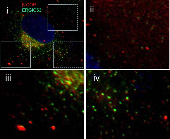
Fig 2.
ERGIC53 and β-COP do not colocalize. Vero cells were fixed, permeabilized, and immunostained for ERGIC53 (green) and β-COP (red). Nuclei were visualized with DAPI (blue). Panel i shows a merged image. Regions of interest taken from the merged image (boxed) are presented at higher magnification (ii to iv) to show separate signals for ERGIC53 and β-COP.

