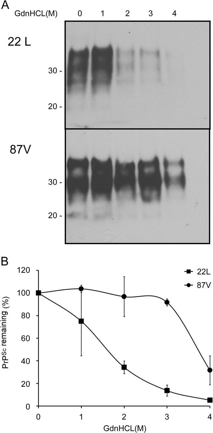
Fig 7.
PrP aggregates in 22L scrapie-infected brain are less stable than those in 87V scrapie-infected brain. (A) Brain homogenates from either 22L or 87V scrapie-infected mice were prepared in PBS at a concentration of 1% (wt/vol) and incubated with increasing concentrations of GdnHCl for 1 h. After the concentration of GdnHCl was adjusted to 0.4 M, samples were centrifuged for 45 min at 17,400 × g. Protein pellets were analyzed by Western blotting using anti-PrP mouse monoclonal antibody 6D11. Molecular mass markers in kilodaltons are indicated on the left. (B) Graph showing the amount of aggregated PrP remaining in the sample following GdnHCl denaturation and centrifugation. The values represent the average values ± standard errors for 2 independent experiments.

