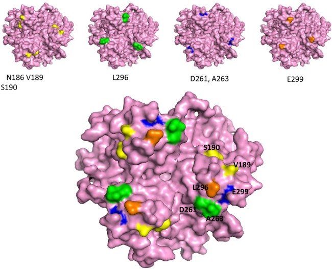
Fig 2.
3D model of the Ad3 fiber knob. The structure is based on that of PDB accession number 1H7Z_A. (Upper) Four critical areas involved in DSG2 binding. The critical residues are shown on the pink isosurface of the trimeric fiber knob. The view is from the top (apical side) facing the receptor. (Lower) All critical residues combined. On the right is an enlargement of the groove after a slight side rotation.

