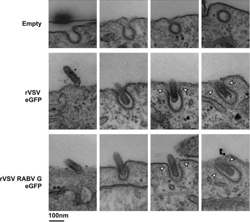
Fig 5.
Visualization of clathrin-dependent uptake of rVSV RABV G by transmission electron microscopy. Electron micrographs show the presumed order of internalization for empty, rVSV eGFP, or rVSV RABV G clathrin-coated pits. BS-C-1 cells were exposed to virus at an MOI of 1,000 for 15 min prior to fixation and processing as outlined in Materials and Methods. Arrowheads highlight sections of the endocytic vesicle lacking electron-dense clathrin.

