
Fig 9.
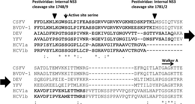
Multiple sequence alignment of NS3 protease/helicase junction. The protein sequences of CSFV (strain Alfort; Protein Information Resource [PIR] accession no. GNWVHC), BVDV-1 (strain NADL; GenBank NP_776267), dengue virus 1 (DEV-1) (strain Brazil/97-11/1997; NP_722463.1), yellow fever virus (YFV) (isolate Ivory Coast/85-82H/1982; Q98803), hepatitis C virus subtype 1a (HCV1a) (isolate 136D_3048_4428_4; ADV92090), and HCV1b (strain MD4-1; AF165051.1) were aligned using clustal omega (version 1.1.0). Positions of the active site serine of the protease domain (vertical arrow) and the Walker A motive of the helicase (horizontal arrow) are indicated. The internal sites of NS3 autocleavages are depicted by inverted triangles. The minimal active protease domain for each virus is marked in bold. Identical, homologous, and similar amino acid residues are indicated below the alignment.