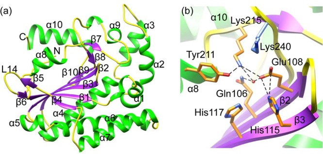
Fig 1.

Overall structure and the nuclease active site. (a) Ribbon diagram of the structure. The α-helices are green, whereas loops and β-stands are yellow and purple, respectively. Helices, β-stands, and loop L14, as well as the N and C termini, are labeled. (b) Nuclease active site showing a network of H bonds (dashed lines in black). The color scheme is the same as in panel a. Side chains of the conserved residues are shown as sticks. The water molecule at the active site is shown as a sphere in red.
