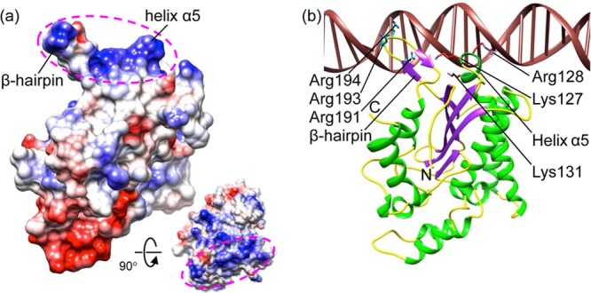
Fig 2.

Putative Ori-recognition site. (a) Electrostatic potential rendition of HBoV-NS1N showing a positively charged surface region mediated by the β-hairpin and helix α5 (magenta ellipsoids). The inset shows a view 90° about the horizontal axis in which the positively charged region faces the reader. (b) Model of HBoV-NS1N (ribbon diagram) binding to dsDNA based on superposition of the AAV-5 Rep nuclease domain structure (PDB code 1RZ9) in the same view as in panel a. Side chains of conserved basic residues are shown as sticks. The N and C termini are labeled. The color scheme is the same as in Fig. 1a. The DNA is in brown.
